64ch / 128slice CT
128
16ch / 32slice CT
32

SUPRIA meets Patient Friendly
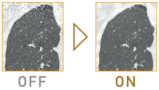
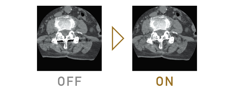
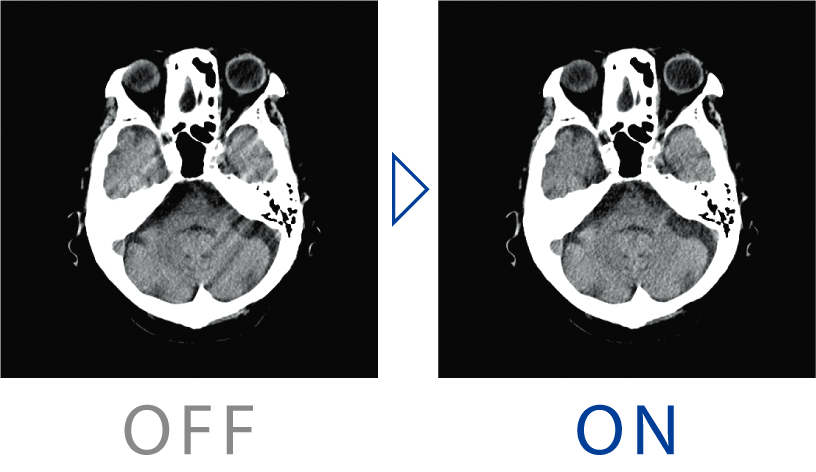
Iterative processing for routine examinations
Iterative Processing, which is useful for dose reduction, requires a large amount of calculation, making it difficult to apply to routine examinations. In "Supria", the image processing unit has been renewed and the processing speed has been improved in order to use iterative Processing (Intelli IP) for routine examinations.
Optimal settings for each facility
Noise reduction strength can be selected from 7 levels. We provide high-quality images by reducing image noise and artifacts with an appropriate exposure dose according to the facility's operation policy.
Low tube voltage scanning
In general, low tube voltage scanning can be expected to increase CT values and improve low-contrast resolution with iodine contrast agents. The noise increased by low tube voltage imaging can be reduced by Intelli IP, also reducing the burden on the patient.
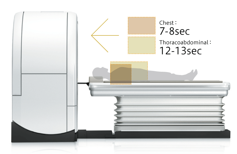
High throughput, high image quality
High performance, such as high-speed rotation, submillimeter slice imaging, powerful X-ray generator and state-of-the-art image reconstruction algorithms, realizes high resolution and high throughput examinations.
SUPRIA meets High Performance
Submilimeter slice imaging for high resolution and high quality images
Supria 32 & 128 shoot high resolution images in a short time based on 0.625mm x 16 inch scanning.
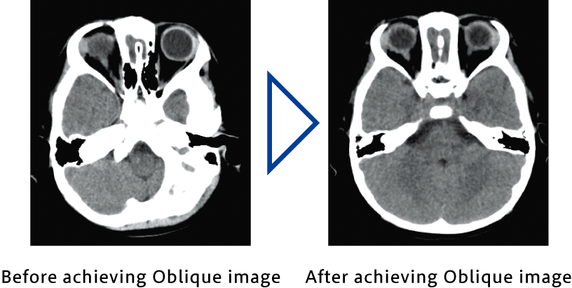
In addition, high resolution and smooth 3D images and MPR images can be achieved by submillimeter slice scanning. Oblique images by MPR can be also achieved after scanning.
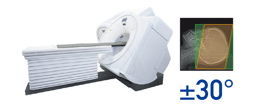
High quality imaging with the gantry tilt
The gantry has a function of tilting within a range of ± 30 degrees, which is possible to reduce exposure to highly radiation sensitive tissues.
In addition, excellent, high-quality images with low contrast can be achieved by normal scan of the head, compared to volume scan. This imaging method takes into account exposure to the patient as well as image quality.
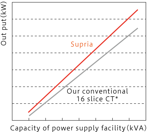
High efficiency powerful X-ray generator
Our technology enabled us to develop a powerful and high efficiency X-ray generator. It achieves sufficient output with a compact power supply facility.
It can also cover heavy load examinations in the X-ray tube, such as wide-area imaging and multi-phase imaging.
SUPRIA meets High Functionality
HiMAR reduces metal artifacts
HiMAR (High Quality Metal Artifact Reduction) adopts unique algorithms for estimating and correcting artifacts based on metal data
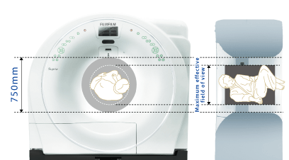
Capable of imaging in various patients' position
With a large bore of 750mm, and a maximum effective field of view that reduces patient anxiety. It is possible to scan the patient in various positions.
ECG Prospective Scanning in synchronization with electrocardiogram
ECG Prospective scanning is a function that scans and achieves image in synchronization with electrocardiographic information. Images achieved by ECG Prospective scanning can be used for calcium scoring analysis*
*1 A 3D workstation equipped with a calcium scoring analysis is required.
Motion Artifact Correction
Body movement can be compensated even after scanning. Even if the patient is out of the effective field of view, such as a patient with a kyphosis, images can be reconstructed without re-scanning in case it is within the maximum effective field of view.
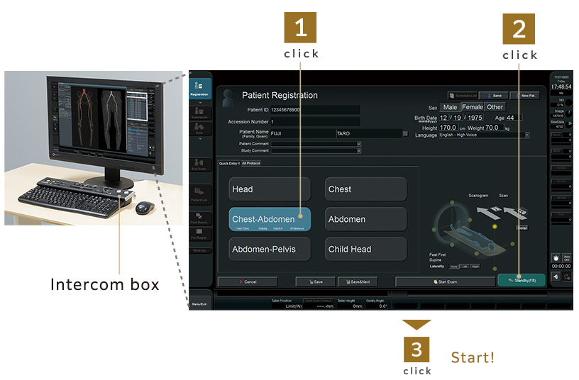
Intuitive operability with Quick Entry
The scan button is located on the intercom box, just above the keyboard, with simply arranged operation buttons, large text, and an easy-to-understand display, which support examinations efficiently.