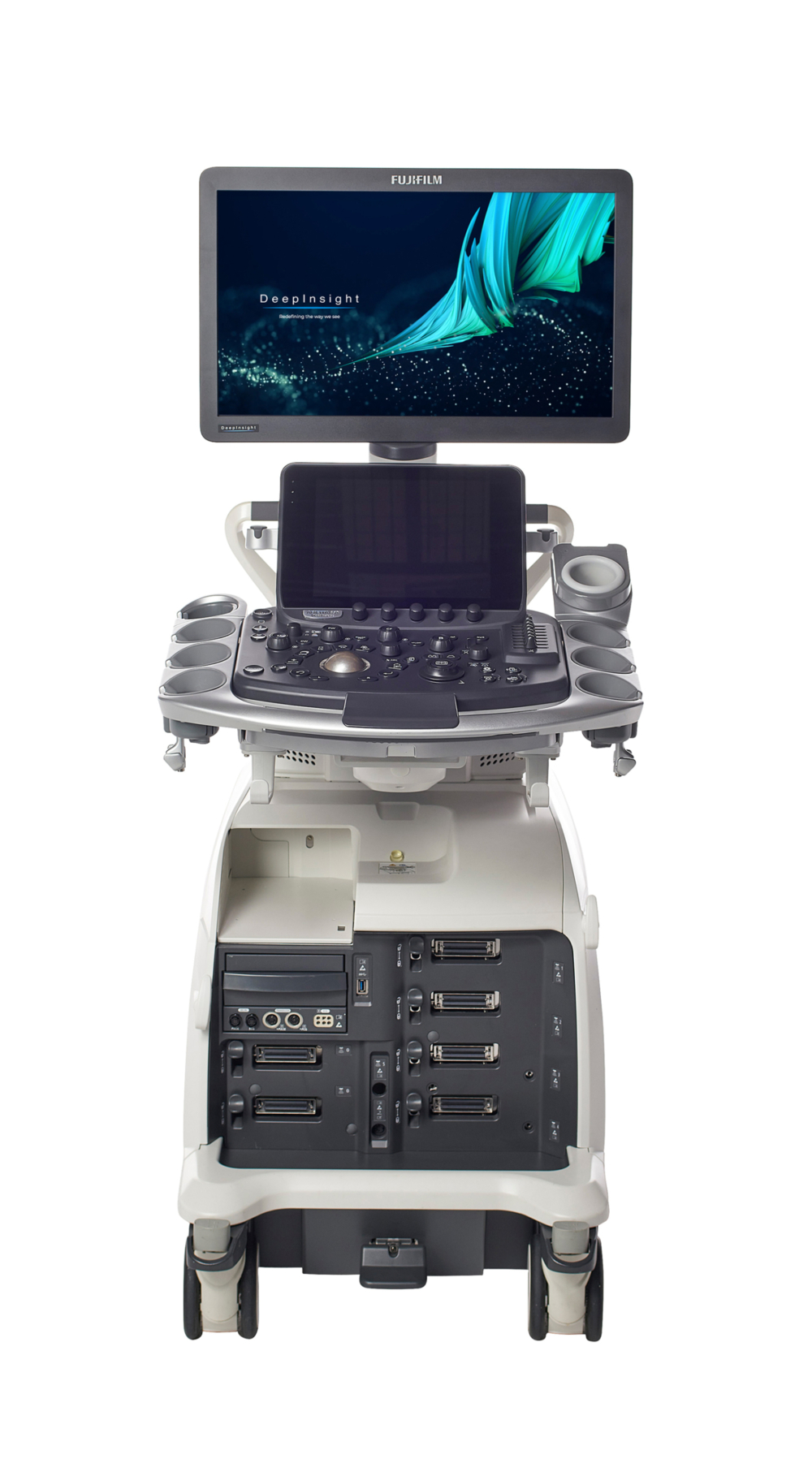
ARIETTA DeepInsight 850
Designed for high expectations
Premium diagnostic ultrasound system which achieves the image quality covering excellent noise reduction, stable penetration, and high spatial resolution with DeepInsight technology.
Greater examination precision, greater comfort, and a wider range of applications are now possible with ultrasound imaging.
Flexibly responding to users' individual needs across the range of clinical disciplines, the ARIETTA 850 DeepInsight brings diagnostic imaging without compromise.

Further refinement of technologies harnessing high quality “sound” gives rise to our highest premium class performance yet.

Designed with sophisticated ergonomics and multiple new tools that streamline your workflow.

An extensive variety of unique applications that deliver new clinical value are accessible across all specialties.
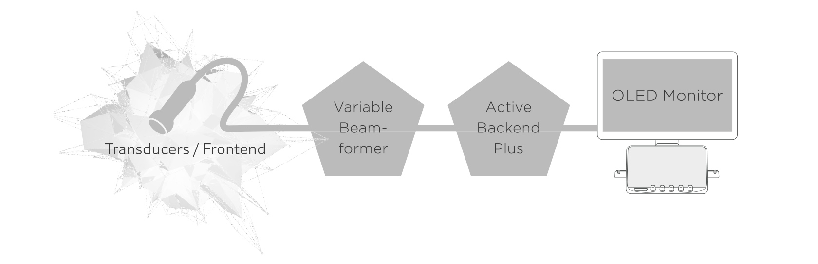
Pure Symphonic Architecture
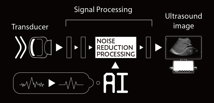
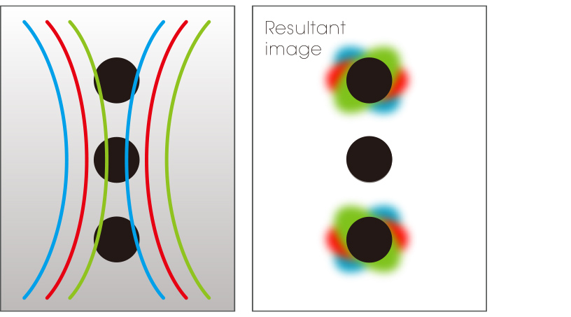
DeepInsight technology, which utilizes AI technology for image enhancement, extracts only the necessary information from a vast amount of data, delivering clearer representations of fine and complex tissue structures that could, until now, have been masked by noise. A more natural representation of the tissue structure is achieved
DeepInsight Technology
The eFocusing technology acquires multiple received beams from a single transmission and combines them to display a single image in real-time. The ARIETTA 850 DeepInsight has evolved the eFocusing technology further by incorporating multiple frequencies to achieve high sensitivity, high contrast, and high spatial resolution.
eFocusing PLUS
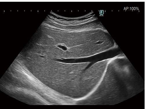
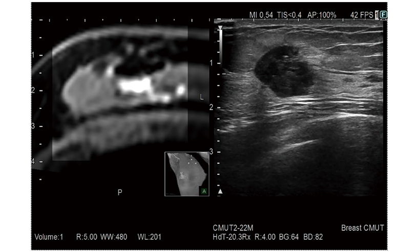
Images with "Clearer Demarcation" are produced by our advanced image processing technology that enhances tissue structure. Delivering stable imaging with less patient dependency.
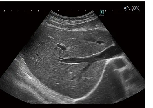
OFF
ON
Carving Imaging
A wide variety of transducers and advanced functions improve accuracy and confidence to deliver therapeutic treatments increasing the curability of lesions.
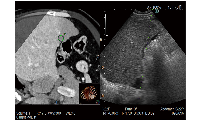
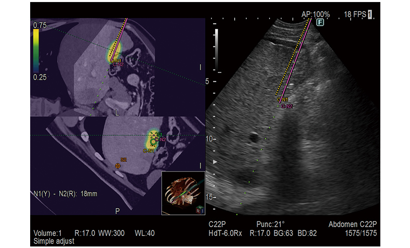
Real-Time Virtual Sonography
RVS is a function which fuses real-time ultrasound imaging with an MPR image created from the previously acquired CT, MRI or ultrasound volume data. It is a complementary technology which allows safer and more accurate treatments such as the detection of tiny lesions which may be difficult to find in an ultrasound examination alone, and the improvement of treatment targeting.
Treatment Support
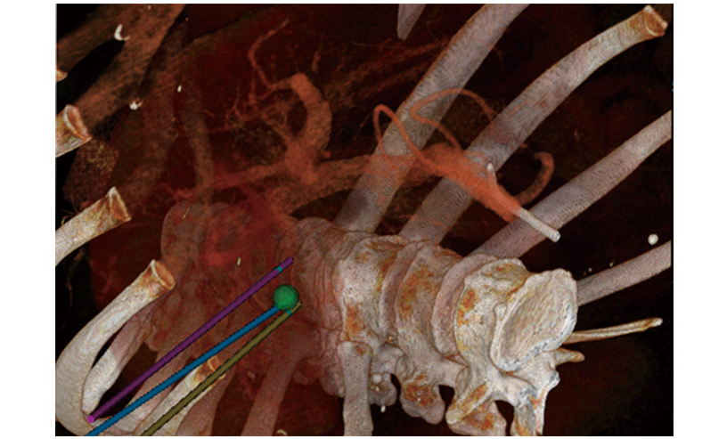
3D Sim-Navigator
A color map superimposed on the CT image simulates the distribution of the electric field (E-field) from the given location of multiple electrodes during RFA treatment. The simulation can be made with different positions of the multiple electrodes or additional ablation to determine the optimal arrangement. This flexibility in planning the needle path can bring significant improvement to the treatment technique.
Provides simulation of single or multiple needle paths during navigation to a target with RVS. The positional relationship between the marked target and needle paths can be assessed in real time using the 3D body mark, reconstructed from the virtual CT volume data, with additional C-plane display orthogonal to the needle path.
E-Field Simulator