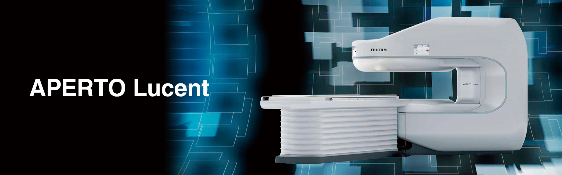
The content on this page is intended to healthcare professionals and equivalents.


0.4T × Open Design

Aperto Lucent offers sophisticated MR imaging through a permanent magnet with 0.4T static field strength together with a compact gantry. 

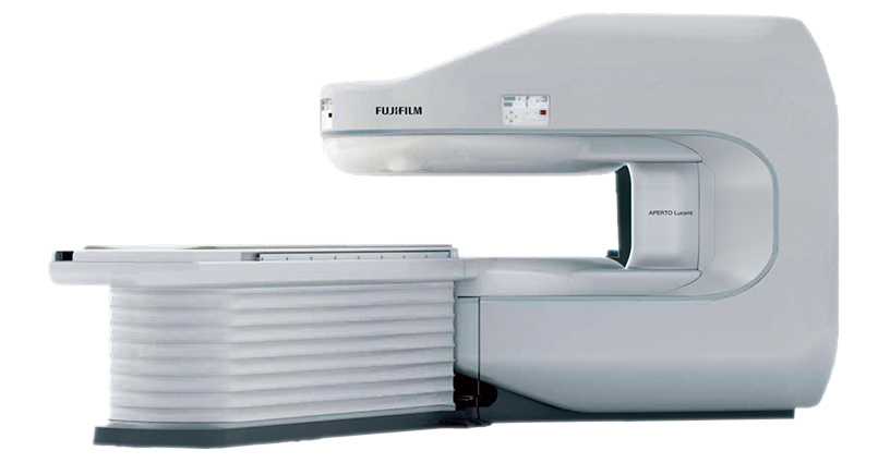
Our technological expertise enabled the design and creation of a single-pillar MRI structure which offers premium open space.

As the only single-pillar MRI system within Our range,the Aperto Lucent offers an expansive, panoramic open aspect designed to reduce patient anxiety and provide a comfortable examination environment.

Floating Table

The lateral slide function allows the floating table to move right and left inside the gantry and the target region can be positioned easily in the centre of the magnetic field.

The table can be lowered to a minimum height of 490 mm, allowing easier accessibility for children and elderly patients.

The 700 mm wide table top offers patients both comfort and a ‘feel-good’ factor, helping to reduce claustrophobia

Open Design

Created to expand space and light, helping to reduce claustrophobia and anxiety

The single-pillar design creates an open examination area which together with the rounded architecture and innovative colour design establishes a secure, calming atmosphere for the patient.


Why choose Open MRI?

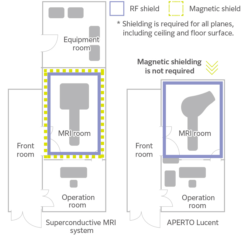
MRI installation usually includes two types of shielding: RF shielding to block any high frequency noise from the outside and magnetic shielding to suppress leakage of the magnetic field from the inside.

However, a permanent magnet MRI system generally does not require any specific magnetic shielding, so the cost of construction is reduced.

Removing many of the construction processes usually associated with superconductive systems, results in faster and easier installation ensuring your Open MRI is up and running in a shorter timeframe.

Ease Of Installation

Aperto Lucent consists of three main units: the gantry, console and power supply system; fewer than its superconductive counterpart.

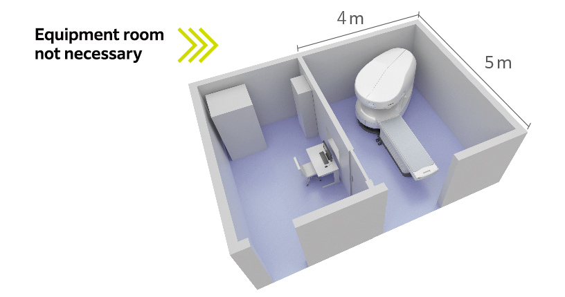
The magnetic field leakage is also kept low, and in turn, the imaging room can be small.

As an equipment room is unnecessary, the overall footprint is reduced and the space saved can be used for other purposes.

Small Footprint

VASC-ASL

Offers non-contrast MR Angiography Technique

VASC-ASL is a non-contrast MRA imaging function that uses 3D BASG (Balanced SARGE) to visualize the blood flow labelled with IR pulses.

This function is used to produce images of portal veins, renal arteries, and upper and lower extremity arteries.  

Reconstruction Imaging

Supports high-definition imaging

This function enables high spatial resolution imaging which result in higher definition images of joint regions required for orthopedic areas.

An image reconstruction matrix of 2048 x 2048 is achieved through the highspeed imaging processor.


Motion Reduction Capability.​

RADAR uses radial scan technology to mitigate motion artefact caused by a patient’s body movement due to voluntary or involuntary motion.

Available with T2WI but also T1WI and FLAIR imaging in any plane and any body region including the head and shoulder joint, which are susceptible to respiration movements and the cervical spine that can be affected by swallowing movements.

RADAR can help reduce repeat scans and improve image quality.

Reduces magnetic field non-uniformity which cannot be corrected with primary shimming.

SuperShim is a technology that increases the uniformity of the static field, which is of paramount importance in MRI.

Non-uniformity in the magnetic field cannot be fully corrected with first order shimming which performs linear correction.

SuperShim is provided to reduce nonuniformity in the magnetic field by enabling high order shimming.


RADAR

SuperShim

High Sensitivity Receiver Coils

Especially effective for images with a small FOV and high spatial resolution

Regions that require a small FOV and high spatial resolution as in orthopedics need higher sensitivity receiver coils.

The solenoid coil adopted in the APERTO Lucent delivers this high sensitivity.

The small diameter coil is tailored to fit the body and the target region easily positioned to the centre of the coil where sensitivity is at the highest.

This is optimal for acquiring images of regions that require a small FOV together with high spatial resolution, as in orthopedics.  


Contact a FUJIFILM Representative: ​ Contact​​ Us

Download Brochure:

* Fujifilm makes no representation that products on this website are commercially available in all countries.

* Approved uses of products vary by country and region.

* Specifications and appearance of products are subject to change without notice.