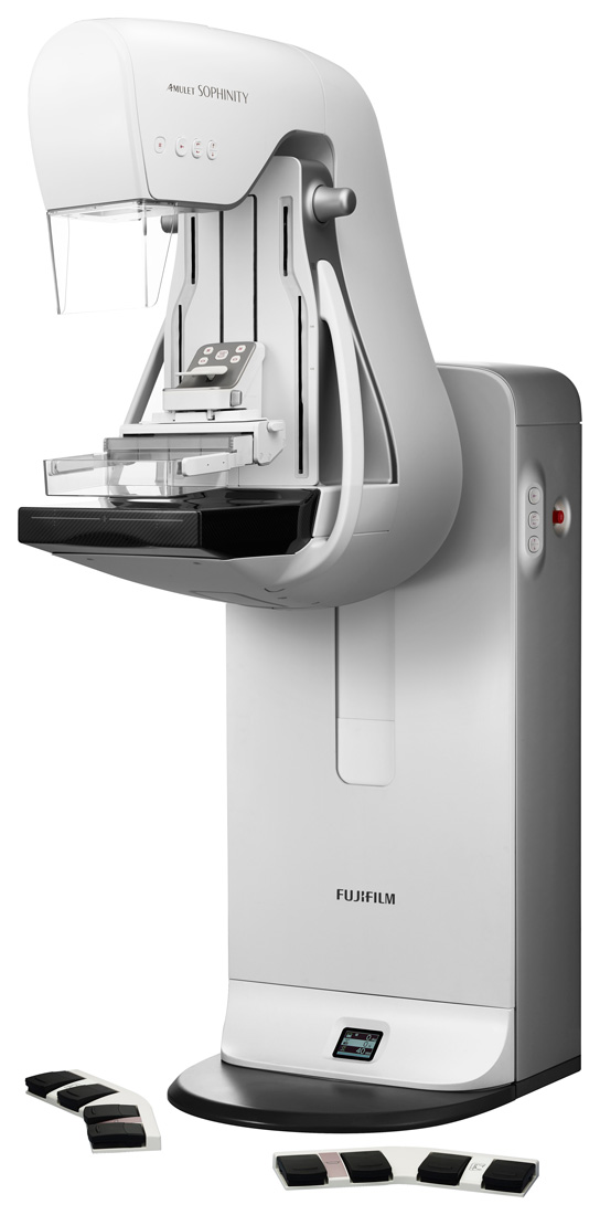

SOPHINITY
Sophisticated features. Human comfort.
Smarter and more comfortable. Pursuing a design concept that is close to the senses of patients, doctors and technologists. This leads to a smooth and relaxed test experience and provides high-quality test results and diagnostic images more reliably. This is the birth of AMULET SOPHINITY, which has refined “gentleness” to meet the needs of all women.
AWS Workstation
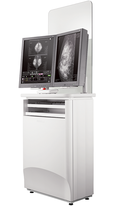
Fully Centralized Acquisition Workstation AWS
- Easy to understand multiple language support
- Integrated X-ray controller allows setting and confirmation of exposure conditions on a single screen.
- Examination screen can be split and switched between 1, 2, or 4 image display.
- Individual images can be immediately output to a PACS, viewer or printer during an examination.
- Density and contrast can be easily adjusted while viewing images.
- Alignment of left and right images can be adjusted both automatically and manually.
Features
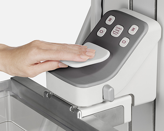
Smooth compression plate operation
Allows fine adjustment of electric compression without removing the line of sight from the breast area during positioning. The compression plate can be operated while confirming the breast condition, contributing to pain reduction for the patient.
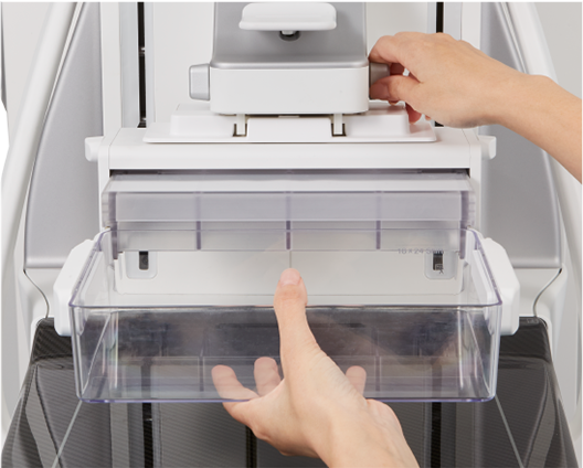
Easy paddle attachment and removal
An array of compression paddles are tailored for the size of the breast and view to be performed. The attachment and removal of the paddles from the gantry is fast and easy for the technologist.
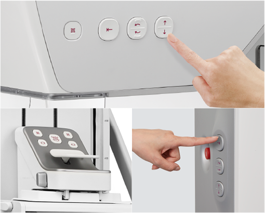
Accessible button layout improves the test flow
Arm rotation and height buttons have been added to the side of the tube head. Easy-to-operate buttons can be selected according to the situation, enabling smoother testing.
High Quality Imaging


TOMOSYNTHESIS
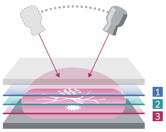
Generates more projection images to reduce artifacts
Continuous tube motion for tomosynthesis sweep and images taken from multiple positions are reconstructed. It can provide images focused on the structures you want to see, further facilitating observation of lesions that are difficult to detect due to overlapping mammary gland structures.

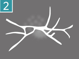
2D Image
Tomography Images
Dynamic Visualization ― Provides high contrast image
It has density/contrast adjustment processing, frequency enhancement processing, and dynamic range compression processing, which allow for automatic adjustment of the amount of dynamic range compression for each image. It recognizes image areas that include the characteristics of the mammary gland and fat areas, and increases the contrast of those areas independently making the density of each constant.

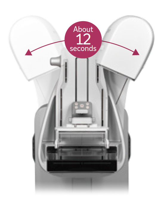
ST (Standard) mode
Sweep angle: ±7.5°
Number of shots: 19
Pixel size: 100/150 m
This mode enables high-speed imaging by reducing the sweep angle and speeding up image readout. The depth of field is deep, and the cine display allows efficient viewing of tomographic images.
HR (High Resolution) mode
Sweep angle: ±20°
Number of shots: 35
Pixel size: 50/100 m
This mode has a larger sweep angle with improved depth resolution. The shallow depth of field allows for a better focus on an area of interest.
FUJIFILM’s medical AI technology brand “REiLI” aims to realize better medical care by combining the image processing technology that FUJIFILM has cultivated to date with cutting-edge AI technologies to provide diagnostic support for doctors in diagnostic imaging and improve workflow efficiency.

FUJIFILM Group supports the Pink Ribbon Campaign
for early detection of breast cancer.